


Dr Sam Bunting 41 Harley Street, London, W1G 8QH
Report 28th August 2023 with reference to Quote SLA-DRS-01
The series of scientific tests was performed by Dr Amy Bowman, working as a scientist for Skin Life Analytics (SLA) under the supervision of Prof Mark Birch-Machin and Jonathan Brookes (Chief Scientific Officer and Chief Executive Officer respectively, Skin Life Analytics).
Background
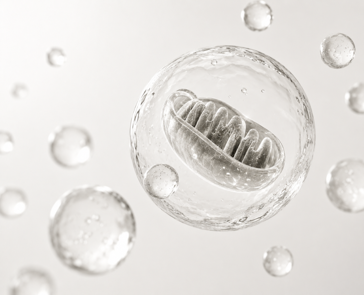
Mitochondria have been shown to be the major site of ROS production in the cell via the electron transport chain (ETC), and mitochondrial DNA (mtDNA) is found in close proximity to the ROS production and has therefore become an established, major target for damage. Many studies have now implicated the key role of mitochondria in the process of skin ageing (summarised in the reviews, references 1 and 2), particularly the role of oxidative stress and mitochondrial dysfunction in the process of skin ageing. Many studies have been conducted to elucidate the mechanism of ageing, and there is continuing evidence that supports the proposal that mitochondria are implicated in both normal ageing and skin photoaging. MtDNA is established as a reliable and sensitive biomarker of UV-induced damage in the skin and the Birch-Machin group has pioneered this over the last 26 years (1-3). This is due to its absence of protective histones, its limited repair mechanisms, and its presence in multiple copies within a cell. Quantitative real-time PCR (qPCR) is used to measure mtDNA damage, based on the principle that qPCR amplification efficiency is decreased in the presence of high levels of UV-induced mtDNA damage.
Methods
In order to determine the viability of the cells following incubation on with various concentrations of Sunflower Shoot Extract (SSE) or niacinamide (NAM) for 24 hours, an MTS cell viability assay was initially used. SSE was tested at up to 5% concentration and NAM up to 5mM. Human dermal fibroblast cells (HDFn cell line) was used to carry out the experiments.
Following the cell viability assays, the optimal concentrations of SSE and NAM were chosen based on concentrations that did not cause a large decrease in viability, as well as previous work. Mitochondrial DNA (mtDNA) damage was determined via real-time qPCR in the presence of these chosen concentrations of SSE or NAM following UV irradiation. To do this, cells were seeded into 35mm dishes and treated with either 1% SSE, 2% SSE, or 1mM NAM Making the Invisible Visible for 24 hours prior to irradiation. SSE or NAM was then removed and replaced with phenol red-free media, and cells were irradiated with a physiological dose equivalent to 2 SED of UVR. SED or Standard Erythemal Doses is not linked to skin type such as minimal erythemal dose or MED. SED is a skin type independent, weighted measurement of sun exposure equivalent to 100 Jm-2, as opposed to MED which is the lowest dose required to produce erythema in an individual. The skin cells used in this study do not exhibit erythema and so MED is not relevant therefore SED represents the unit of dose (11 and htps://www.ncbi.nlm.nih.gov/pmc/articles/PMC6801664/ ). The lamps used are Cleo performance 100W-R, IsoLde, Stutgart, Germany which are 6 feet lamps housed in custom made equipment. This was used in our action spectrum study published in the J Invest Dermatol. where the exact Cleo lamps and housing were used. The paper reference for the lamps is reference 4 below, Latimer et al., 2015. The full spectrum of the Cleo was used to calculate the 2 SEDs.
The 100% covered dishes were wrapped in aluminium foil to represent complete protection (negative control). DNA was extracted following UV using the manufacturer’s instructions using a QIAamp DNA Mini Kit (Qiagen, UK). Total DNA concentration was determined using a NanoDrop ND2000 (ThermoFisher, UK). The levels of mtDNA damage within the cells were determined via real-time qPCR using a QuantStudio™ 3 Real-Time PCR System (Applied Biosystems, UK). MtDNA damage is an established biomarker of UV damage in skin, and the level of UV-induced damage within the mtDNA was determined via real-time qPCR using Skin Life analytics proprietary assays. MtDNA damage is expressed as a Ct value, where a 1 Ct difference is equivalent to a 2-fold difference in damage. An 83bp assay was also performed for each condition to determine the relative amount of mtDNA present (ref 5). This to ensure that the level of mtDNA analysed was the same across all samples and can therefore be used to determine the overall amount of mtDNA content per sample (5). Three biological repeats were performed for each condition, with the PCR run in triplicate for each sample.
Results
1. Cell Viability
Cell viability measurements was determined to ascertain the highest concentration of product (Sunflower Shoot Extract (SSE) or Niacinamide (NAM)that can be tolerated by the cultured living human skin cells (human skin dermal fibroblasts). This was determined using the MTS cell viability assay.
Figure 1 shows the MTS cell viability results in the presence of various concentrations of either SSE or NAM.
As can be seen in Figure 1a, SSE did not cause a decrease in cell viability even up to a concentration of 5%. This suggests that SSE is not toxic to the human dermal fibroblast (HDFn) cell line used. Following discussions with Dr Bunting and colleagues, concentrations of 1% and 2% SSE were chosen as the optimal concentrations to test for their ability to provide UV protection against mtDNA damage.
As seen in Figure 1b, NAM only decreased cell viability to 90% at 1mM, so this concentration was chosen to be used in future experiments, based on previous work in our lab showing positive effects on mitochondrial function at this concentration (ref 6).

Figure 1. HDFn fibroblast skin cell viability in the presence of SSE or NAM was determined via an MTS assay, in the presence of various concentrations of either SSE (a) or NAM (b) for 24 hours. SSE did not cause a decrease in cell viability up to 5%, and NAM showed a 90% viability at 1mM (within the acceptable concentration). 3 biological repeats were performed, with 8 technical repeats per condition per run (each column = 24 data points).
2. Mitochondrial DNA (MtDNA) protection:
Using Skin Life analytics proprietary assays, the SSE and NAM products were evaluated for their efficacy in atenuating the damage to mitochondrial DNA (MtDNA) in a monolayer culture of human dermal fibroblast skin cells using a physiological UVR dose.
Figure 2 shows the levels of UV-induced mtDNA damage in skin cells following 24 hour treatment with 1% SSE, 2% SSE, or 1mM NAM, followed by UV irradiation at 2 SED. MtDNA damage is expressed as a Ct value, as a combined result of 3 biological repeats (with each biological repeat performed in triplicate on the PCR assay i.e the technical repeat). ). The higher the Ct value on the y-axis, the greater is the amount of MtDNA damage. As can be seen in Figure 2, mtDNA damage was much greater when the cells were exposed to 2 SED UV (100% exposed, i.e .positive control) as compared to when the cells were completely foilcovered as expected (P<0.0001***). Each 1 Ct difference represents a 2-fold difference in damage.
SSE at a concentration of 1% showed UV protection of mtDNA compared to the 100% exposed cells, as seen by decreasing the level of mtDNA damage by approximately 0.4 Cts. This equates to approximately 1.3 fold less damage in the cells with 1% SSE, or 22% less damage. This difference in damage between 1% SSE and the 100% exposed cells was statistically significant (P= 0.029*), suggesting that 1% SSE is providing UV protection for skin cells.
SSE at 2% also provided mtDNA damage protection against UV compared to the 100% exposed cells, decreasing the level of mtDNA damage by approximately 0.6 Cts. This is approximately 1.5 fold less damage, or 34% less damage. This difference in damage was also statistically significant (P= 0.012*), suggesting that 2% SSE is providing UV protection, with a slightly higher level of mtDNA damage protection than 1% SSE.
In contrast, under the same experimental conditions, 1mM NAM did not appear to provide any mtDNA damage protection against UV irradiation following pre-treatment for 24 hours (P= 0.511, not statistically different). This may be expected based on previous work in our laboratory, which has found that the beneficial effects of 1mM NAM on mitochondrial parameters requires up to 7 days of pre-incubation with human skin fibroblasts (ref. 6). We used NAM at 24 hours to ensure the same conditions as for the SSE, with our results suggesting that 1% and 2% SSE provide higher levels of UV protection than NAM under the same conditions.
The effects of the SSE treatment alone did not appear to be the cause of the observed decrease in mtDNA damage following UV irradiation, as those cells that were treated with 1% or 2% SSE but covered with foil during the irradiation did not show any decrease in damage compared to the media control covered with foil (results not shown).
An 83bp assay was also performed to ensure that the same amount of mtDNA was present per sample. This tells us that the protection of UV-induced MtDNA damage provided by 1% and 2% SSE were not due to differences in mtDNA content.

Figure 2. UV-induced mtDNA damage levels in skin cells following 24 hour treatment with SSE or NAM. Real-time qPCR was used to determine the levels of mtDNA damage in human dermal fibroblast cells (HDFn cells), following UV irradiation. Cells were treated for 24 hours prior to irradiation with either 1% SSE, 2% SSE, or 1mM NAM, which were removed before irradiation. Cells were irradiated with 2 SED UV light, and mtDNA damage determined via real-time qPCR. MtDNA damage is expressed in the figure as a Ct value (where for example, a 1 Ct difference is equivalent to a 2-fold difference in damage). The higher the Ct value, the greater is the amount of MtDNA damage. Both 1% SSE and 2% SSE provide a statistically significant degree of UV protection. Three biological repeats were performed, each representing the average of three technical repeats on the qPCR plate (therefore each column represents 9 individual data points). Damage was decreased significantly from the 100% exposed sample following incubation with 1% SSE (P= 0.029*) and 2% SSE (P= 0.012*). There was no significant change with 1mM NAM (P= 0.511).
Table 1 shows the raw data for Figure 2, and Table 2 shows the statistical analysis results.
Table 1

Table 1. Raw data for Figure 2. Table 1 shows the raw data acquired for Figure 2. Ct values are shown as determined via qPCR, representing the level of mtDNA damage. The higher the value, the more mtDNA damage is present. Each repeat value represents the average of three technical repeats on the qPCR plate, for the three biological repeats.
Table 2

Table 2. Statistical analysis of data. Statistical analysis of the Ct values obtained in Figure 2 was performed using an unpaired t-test, for treatments compared to the 100% exposed sample. Significant differences were seen between the 100% exposed sample and the 1% SSE, 2% SSE, and 100% covered with foil cells.
Summary
This set of experiments provide biological evidence in cultured human skin cells that clearly demonstrate the potency of protection by SSE against UV-induced mtDNA damage which is an established marker of UVR and sun exposure damage in human skin as well as being a marker of skin aging. There is a clear protection provided by SSE against MtDNA damage which is statistically significant for both the 1% and 2% SSE. In contrast 1mM NAM under the same experimental conditions did not provide statistically significant MtDNA protection This is certainly an exciting biological finding as the outcomes show the provided SSE protects against damage to the DNA housed inside the batteries of the cell. This will help in the battle against skin fatigue and help in the process of boosting vibrant energetic skin and of course being kind to your skin DNA.
References
- Naidoo K; Hanna R; Birch-Machin MA (corresponding author). What is the role of mitochondrial dysfunction in skin photoageing? Exp Derm, 2018, 27, 124-128. doi: 10.1111/exd.13476
- Stout R and Birch-Machin MA, Mitochondria’s Role in Skin Ageing. Special Issue "Mitochondrial Dysfunction in Ageing and Diseases of Ageing". Biology 2019, May 11;8(2). pii: E29. doi: 10.3390/biology8020029.
- Bowman, A. and M.A. Birch-Machin, Age-Dependent Decrease of Mitochondrial Complex II Activity in Human Skin Fibroblasts. J Invest Dermatol, 2016. 136(5): p. 912-919.
- Latimer J, Matts P, Diffey B, Lloyd J and Birch-Machin MA (corresponding author). Determination of the action spectrum of UVR-induced mitochondrial DNA damage in human skin cells.. J Invest Dermatol. 2015, 135 2512-2518.
- Hanna R, Crowther J, Bulsara P, Wing X, Moore, D and Birch-Machin MA (corresponding author). Optimised detection of mitochondrial DNA strand breaks. Mitochondrion 2019, 46, 172-178.
- Oblong, J. E., Bowman, A., Rovito, H. A., Jarrold, B. B., Sherrill, J. D., Black, M. R., Nelson, G., Kimball, A. B. and Birch-Machin, M. A. Metabolic dysfunction in human skin: Restoration of mitochondrial integrity and metabolic output by nicotinamide (niacinamide) in primary dermal fibroblasts from older aged donors. Aging Cell 2020, 19(10), p. e13248.